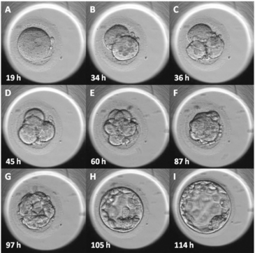
Time-lapse embryo imaging is one of the most discussed technological advances in IVF over the past decade. The idea is compelling: instead of removing embryos from the incubator once or twice a day to assess their development under a microscope, a camera inside the incubator captures thousands of images automatically, creating a continuous record of how each embryo divides and grows.
At CEGYR, we have evaluated this technology carefully. Here is what the evidence actually shows — and what it doesn’t.
How time-lapse systems work
Time-lapse incubators (such as the EmbryoScope) contain a built-in camera that captures images every 10 to 20 minutes. These images are combined into a short video showing days of embryo development compressed into minutes. The result is a detailed picture of each embryo’s division timing, cell number, symmetry, and the appearance of abnormalities that might be missed during a single observation window.
The two proposed benefits — and the evidence behind them
1. Undisturbed development
The most intuitive benefit is that embryos never have to leave the incubator. Traditional assessment requires briefly removing the dish and exposing embryos to changes in temperature, CO₂, and handling. Time-lapse systems eliminate this disruption entirely.
Whether this translates into meaningfully better outcomes is a legitimate hypothesis — but the randomized controlled trials conducted to date have not shown a consistent, significant improvement in pregnancy rates from this effect alone.
2. Better selection through morphokinetics
The second claimed benefit is more sophisticated: by analyzing the exact timing of each cell division (a field called morphokinetics), software algorithms attempt to predict which embryos have the highest developmental potential. If the algorithm correctly identifies the “best” embryo to transfer, time-to-pregnancy could theoretically be shortened.
Here, the evidence is mixed. Some studies show modest improvements in outcomes when morphokinetic criteria are used for embryo selection. Others show no significant difference compared to conventional assessment by experienced embryologists. The algorithms require large, well-calibrated datasets to perform reliably — and performance can vary between clinics.
What we conclude at CEGYR
Time-lapse imaging is a genuinely interesting technology. It adds information, and the principle of minimizing embryo disturbance is sound. But it is not a substitute for a high-quality embryology laboratory, experienced embryologists, and evidence-based stimulation protocols — which remain the primary drivers of IVF success.
We use time-lapse as a complementary tool, not a standalone differentiator. When patients ask whether a clinic’s “time-lapse system” means better results, the honest answer is: it depends on the entire laboratory ecosystem around it.
Questions to ask your clinic
If a clinic emphasizes time-lapse imaging as a key selling point, it is worth asking:
- What are their published or audited pregnancy rates — not marketing claims, but real data?
- Do they use the morphokinetic data to make transfer decisions, and if so, based on which validated algorithm?
- What is the experience level of their embryology team?
Technology improves outcomes when it is embedded in a culture of evidence and excellence. It does not replace them.